
By Émile Lemoine
“Epilepsy and autism are incredible models to study brain networks.”. Professor Boris Bernhardt, founder and Principal Investigator of the MICA (Montreal Imaging and Connectome Analysis) Lab, at the McConnell Brain Imaging Center of the Montreal Neurological Institute, dedicates his research to these diseases that offer a window into brain development, functional networks, cortical microstructure, and high-level cognitive functions like memory, attention and self-generated thoughts.

Boris Bernhardt studied cognitive science in Germany, with an interest in computational neurosciences. An internship opportunity during his early studies at McGill University had a lasting effect on his career. Later, he would pursue his Ph.D. at McGill, under the supervision of Dr Neda Bernasconi, where his thesis focused on cortical thickness in temporal lobe epilepsy. After working both at McGill and at the Planck Institute of Human Cognitive and Brain Sciences in Germany as a postdoc, he settled down in Montreal and founded the MICA in 2015, where he and his team develop data-driven solutions to fundamental questions in neuroscience.
From microstructure to function in temporal lobe epilepsy
Drug-resistant epilepsy is a neurosurgical condition affecting one third of epilepsy patients, with seizures most often originating in the temporal lobe1. While classical temporal lobe epilepsy models described a well-localized seizure focus with propagation to the surrounding cortex, neuroscientists now agree that the network underlying this disease is much more distributed throughout the brain. Many studies attempt to describe these networks by mean of neurophysiological monitoring or resting-state brain mapping. Meanwhile, brain specimens of epileptic patients have long manifested alterations at the microscopic scale: neuronal loss, gliosis (reactive increase in glial cell count secondary to inflammation), and cortical dysplasia (congenital local malformation of neuronal architecture). With advanced analytical techniques, MRI can reveal epileptic markers in vivo: cortical thickness, white matter connection patterns, grey-white matter interface, volumetric analysis of specific brain regions, and many others. Unfortunately, these changes exhibit a much more widespread distribution, to the extent that we cannot tell if they are responsible for the disease, or its consequence.

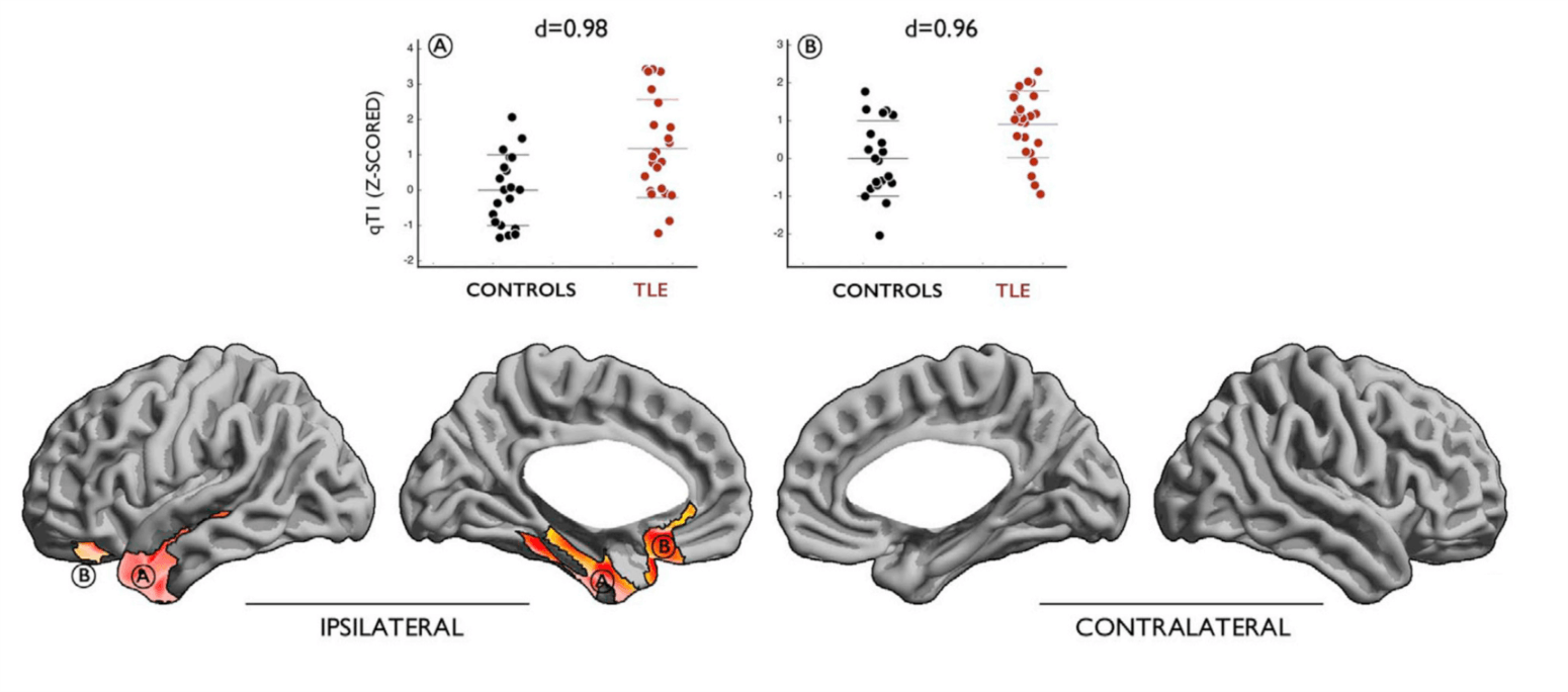
In their article Preferential susceptibility of limbic cortices to microstructural damage in temporal lobe epilepsy: A quantitative T1 mapping study2, published in 2018 in NeuroImage, Pr. Bernhardt and his co-authors argue that quantitative MRI—mapping the time it takes for tissue magnetization to recover from an initial radiofrequency excitation back to its initial state—could help characterize epileptic brains better than previous techniques. “Traditionally, specimens from epileptic patients were analyzed post-mortem or after surgical excision, and only showed non-specific alterations, such as gliosis or neuronal loss. Sometimes, however, pathologists would note hints of abnormalities in the cortical architecture itself.” This is important, and is at the heart of the article: how does epilepsy emerges from the organization of the neurons in the cerebral cortex? To date, MRI studies have not offered a clear answer: “Many researchers, including myself, have studied morphological changes in the brain of epileptic patients—cortical thickness and volumetric analysis, for example. However, these changes are widespread across the brain, whereas temporal lobe epilepsy is a lateralized and localized disorder, with origins in the limbic system. These morphological markers are therefore not specific to the pathology underlying temporal lobe epilepsy and may be markers of damages secondary to medication or neurotoxicity from the seizures.”
The study, which started in 2014, consisted in mapping quantitative T1 relaxation time (qT1) at 3 Tesla in patients with temporal lobe epilepsy, and compare these maps with healthy controls. The results demonstrate that qT1 distribution across the cortex is effectively altered in epileptic patients and can identify the side of the seizure focus with an accuracy over 90%. But more importantly, these changes occur preferentially in the limbic cortex, and remain significant even after controlling for other MRI markers such as cortical thickness and grey-white matter interface blurring. “We were excited when we saw the results. However, there remains a long way before this marker is used in the clinical setting. The interpretation of qT1 is still speculative; we have good reasons to think it may inform us of myelinated fibers, but there may be other things. There is never a one-to-one mapping between MRI and nature!” When asked if the team will pursue the study of quantitative MRI in epileptic patients, Pr. Bernhardt affirms that he is currently organizing a project in collaboration with neuroscientists in Vancouver, BC: “We are investigating quantitative times in kids, both with and without epilepsy syndromes, to give us a sense of the neurodevelopmental aspect of quantitative MRI changes in epilepsy.”
Teamwork
Collaborations between laboratories are a necessity when studying diseases like epilepsy. The sparsity of high-quality MRI data is a major hurdle, especially when increasingly powerful computational techniques and machine learning algorithms rely on large datasets to achieve their full potential. “Not only is more data better to train machine learning models, but we need even more data to ensure that the models generalize well across different centers and cohorts.” In many fields, the free sharing of data and computer code is becoming a popular way to overcome these difficulties and accelerate the development and evaluation of new technologies. The MICA is an active participant of the Open-Science community. “My first investment as a young principal investigator was to acquire Connectome-in-a-Box from the Human Connectome Project. In my career, I have benefited maximally from people sharing their code and data.” Now, it is his turn share to the work of his team with other investigators.

Since its foundation, the lab has seasoned many students from diverse backgrounds: engineering, neuroscience, computer science… all sharing a common passion for the brain and data analysis. “It really is a mixed bag of people, all very talented and motivated and fun to work with.” He reassures that you do not have to be born writing computer code to work with him: “They all engage in quantitative analysis at some point, but the dedication is more important than a strong programming background. Everyone can bring something to the team, and that is my job, as supervisor, to put everyone in the right niche.” How hard it is for him to enforce code sharing practices into its student? “Most of the times, it comes from them! They see how much it accelerates their own research when they use the tools others have developed before them. This results in a real synergy between students.”
Initiatives such as the Human Connectome Project have immensely contributed to the popularization of neuroimaging research. However, disease-specific projects of that scale are still emerging. “Unfortunately, in epilepsy, large scale data-sharing initiatives do not really exist. Except for ENIGMA.” The ENIGMA Consortium regroups leaders in the fields of neuroimaging, neurogenetics, neurology and psychiatry from all-over the world to combine their data and minds in order to discover solutions to problems that individual research groups with small cohort-sizes cannot tackle. ENIGMA-Epilepsy was launched in 2015. “ENIGMA-Epilepsy is a breakthrough for reproducible epilepsy research and is the first step in standardized data sharing of epilepsy neuroimaging. Right now, the consortium is aggregating data from 21 sites, and the first paper was published last year. We are talking about 2,000 patients and over 1,000 controls.” Indeed, the article published in Brain in 2018 presents findings from “the largest neuroimaging study of epilepsy to date”, with 2149 epilepsy patients from 24 research centers in 14 different countries3.

Pr. Bernhardt worked on this paper and plans to keep this collaboration active on future projects. “Up to this point, centers could submit their data to a coordinating center, which performs a meta-analysis on the images. The next step is to make available the preprocessed features extracted from the images, to perform mega-analyses. The ultimate goal would be to share the real and anonymized MRI data, just like the Human Connectome Project.”
Between epilepsy and autism
Beside epilepsy, researchers at the MICA study another neurodevelopmental disorder: autism spectrum disorder (ASD). “Initially, I was more interested in the co-occurrence between autism and epilepsy. But the overlap is not so prevalent, as these diseases are highly heterogenous, so I have come to study both diseases as separate entities.” While these two disordered have developmental origins, findings in one area do not necessarily translate into the other: “Temporal lobe epilepsy and ASD have characteristic neuroimaging findings, and commonly exhibit comorbid, high-level cognitive abnormalities, such as disorders of memory or attention. In ASD, however, the neuroimaging abnormalities have no clear localization, and tend to be more diffuse.” The recruitment is also different in both cases: “For autism research, the patients we study are children.” Indeed, the effect size of ASD is larger earlier in life; brain overgrowth, increased cortical thickness and other markers tend to normalize with age, and so do the clinical manifestations. “It is funny because in epilepsy, we look more at the effect of the disease towards adulthood, whereas for autism, we focus on the developmental aspect, and how the brain changes through time.”
For future years, Pr. Bernhardt has exciting ambitions. After rapid expansion of the lab, he now invests in large-scale collaborative initiatives, such as the ENIGMA-Epilepsy project. With his team and other research groups, he envisions to further investigate the links between cortical microstructure and functional connectivity, both in diseased and healthy patients. “The research now is pretty much centered around three themes: 1) investigation of brain structure-function and large-scale cortical organization of cognitive circuits, especially the limbic network, 2) combination of microstructure imaging with connectomes and how they are both affected by brain lesions, and 3) understand the development of these circuits and how it could contribute to diseases such as epilepsy and autism.” With researchers as passionate as Pr. Bernhardt on their heels, epilepsy and autism better find a good hideout!